Muscle Physiology


Skeletal or striated muscle cells are multi-nucleated and under voluntary control. They are responsible for producing movement, generating heat, maintaining posture, and stabilizing joints.

All skeletal muscles exhibit four characteristics:
  • Excitability - the ability to receive and respond to a stimulus

  • Contractility - the ability to shorten

  • Extensibility - the ability to stretch or lengthen beyond resting length

  • Elasticity - the ability to recoil and resume its resting length

Skeletal Muscle Organization
Muscle cells are covered with endomysium and then bundled into groups called fasicles. The fasicles are covered by the perimysium, bundled together and covered by the epimysium. Tendons connect the muscle to the bone. All connective tissue coverings (endomysium, perimysium, epimysium) are continuous with the tendon.

Skeletal Muscle Cell Anatomy
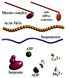


  • sarcolemma - the plasma membrane of a muscle cell

  • sarcoplasmic reticulum - the endoplasmic reticulum of a muscle cell

  • sarcomere - myofibril region between two successive Z discs

  • A band - region of a sarcomere where the myosin (thick) myofilaments are found

  • I band - region of a sarcomere where the actin (thin) myofilaments are found

  • M line - center of the H zone

  • Z disc - anchors the actin (thin) filaments and connects the myofibrils

  • H zone - area within the A band where actin and myosin filaments do not overlap


  • Filament Structure
    There are three filament types found within a sarcomere: myosin, actin, and elastic.

    • Myosin - thick filaments
      - 2 interwoven protein chains
      - 2 globular heads terminate the chain known as cross bridges
      - actin and ATP binding sites are present

    • Actin - long, thin filaments
      - helical structure with active binding sites for myosin heads
      - contains tropomyosin - a protein to help stiffen the actin core; in relaxed muscles they block myosin binding sites
      - contains troponin - a three protein complex
      Protein 1 binds to actin
      Protein 2 binds to tropomyosin and helps to move it
      Protein 3 binds to calcium ions

    • Elastic Filaments
      - holds the myosin (thick filaments) in place
      - helps the muscle stretch and recoil

    Steps in Excitation-Contraction Coupling
    &
    The Sliding Filament Theory of Contraction

    The following events occur from the time of action potential to mechanical activity.
    1. An action potential fires at the neuromuscular junction.
    2. Neurotransmitter is released from the axonal terminal, diffuses across the synaptic cleft, and attaches to receptors on the sarcolemma.
    3. The action potential travels down the sarcolemma and down the T-tubules.
    4. Calcium ions are released from the sarcoplasmic reticulum and they bind to troponin. This causes a change in shape in troponin, removing the blocking action of tropomyosin exposing the actin binding sites. 5. Myosin cross bridges to become activated and attach to the actin binding sites.
    6. The myosin head changes shape and pulls on the actin sliding it to the center of the sarcomere. ATP energy is used to accomplish this step.
    7. A new ATP molecule binds to the myosin head. The cross bridge detaches from the actin. The new ATP molecule has caused the myosin head to change shape and the head can now reattach to the actin to repeat the process in step 6. 8. Contraction continues until the calcium ions are removed by active transport back into the sarcoplasmic reticulum.
    9. Once the calcium is removed from troponin, tropomyosin moves to its original shape and blocks the actin active sites causing muscle relaxation.


    There are 2 main categories of contractions: isotonic and isometric.
    Isotonic Contractions occur when the muscle changes length and results in decreasing the angle at the joint to move the load. There are two types of isotonic contraction.
  • Concentric contractions - the muscle contracts as it shortens
  • Eccentric contractions - the muscle contracts as it lengthens

  • Isometric Contractions occur when the muscle does not shorten or lengthen, yet tension continues to increase.

    There are many ways to classify muscle fibers. Two major characteristics are used in muscle fiber classification, the speed of contraction and the dominant way the muscle fiber produces ATP.

    The speed of contraction of a muscle fiber is directly related to how fast cells are able to split ATP to release the molecules energy for use. Fibers that have efficient ATPase are known as fast fibers. Fibers that have slow ATPase are known as slow fibers.

    ATP generation is essential for muscle fibers. If a muscle fiber uses oxygen or aerobic pathways, they are called oxidative fibers. By contrast, glycolytic fibers use anaerobic patways like glycolysis.

    Muscle Animation I
    Muscle Animation II