
|
|
|
|
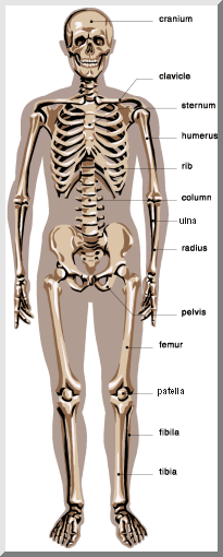
The skeleton is composed of bones, cartilages, joints, and ligaments. There are 206 bones in the human skeleton which are divided into the axial and appendicular divisions. The axial skeleton is comprised of the skull, vertebral column, and rib cage. The four appendages, the pectoral girdle and the pelvic girdle make up the appendicular skeleton. |
|
|
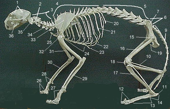
The feline skeleton is also divided into two divisions: the axial and appendicular skeleton.
|
|
| The Facial Bones Include:
1. the nasal bones
2. the maxilla bones
3. the zygomatic bones
4. the mandible
5. the lacrimal bones
6. the palantine bones
7. the inferior conchae bones
8. the vomer bone
|  |
| Key to the Anterior View of the Skull:
1. frontal bone
2. supra-orbital foramen
3. orbit (orbital cavity)
4. superior orbital fissure
5. inferior orbital fissure
6. zygomatic bone
7. infra-orbital foramen
8. maxilla
9. mandible
10. mental foramen
11. incisive fossa
12. symphysis
13. vomer
14. inferior nasal concha
15. middle nasal concha
16. perpendicular plate of ethmoid
17. nasal bone
18. lacrimal bone |
| The Cranial Bones Include:
1. frontal bone
2. parietal bones (paired)
3. temporal bones (paired)
4. occipital bone
5. sphenoid bone
6. ethmoid bone |  |
| Key to the Lateral View of the Skull:
1. Parietal Bone
2. Coronal Suture
3. Frontal Bone
4. Nasal Bone
5. Vomer
6. Lacrimal Bone
7. Orbital Part of Ethmoid
8. Zygomatic Bone
9. Maxilla
10. Body of Mandible
11. Ramus of Mandible
12. Coronoid Process
13. Mandibular Condyle
14. Mental Foramen
15. Styloid Process
16. External Acoustic Meatus
17. Mastoid Process
18. Zygomatic Process
19. Temporal Bone
20. Greater Wing of Sphenoid
21. Inferior Temporal Line
22. Superior Temporal Line
23. Squamosal Suture
24. Lambdoidal Suture
25. Occipital Bone
|
 |
 |
 |  |
 |
 |
 |
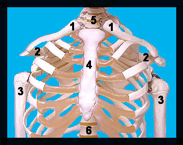
| The bony thorax consists of the thoracic vertebrae, the ribs, the sternum, and the costal cartilages. |
| The sternum or breastbone includes the fusion of three bones: the manubrium, the body, and the xiphoid process. |  The Sternum: 1. Jugular Notch 2. Manubrium 3. Sternal Angle 4. Body of Sternum 5. Xiphoid Process |
 | There are 12 pairs of ribs. Seven pairs are true ribs which attach directly to the sternum. The remaining 5 pairs are false ribs attaching indirectly to the sternum. Pairs numbered 11 and 12 are called floating ribs because they lack an anterior attachment site. |
| The Appendicular Skeleton |
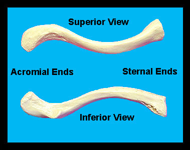
| The pectoral girdle is comprised of two bones: the clavicle and the scapula. The pectoral girdle serves to attach the upper limbs to the body. |
 |
 |
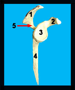 The Scapula (lateral view): 1. Acromion process 2. Coracoid process 3. Glenoid cavity 4. Lateral (axillary) border 5. Spine |
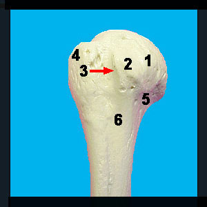 The Humerus (anterior-proximal end): 1. Head of the humerus 2. Lesser tubercle 3. Intertubercular groove 4. Greater tubercle 5. Anatomical neck 6. Surgical neck | 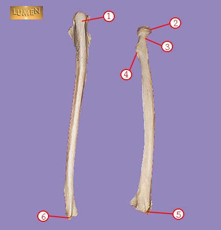 The Radius (right) and Ulna (left)(posterior view): 1. Posterior aspect of the olecranon process 2. Radial head 3. Neck of the radius 4. Radial Tuberosity 5. Styloid process of the radius 6. Styloid process of the ulna |
| The pelvic girdle attaches the lower limbs to the axial skeleton, protects the organs of the pelvis, and transmits the weight of the upper body to the lower limbs. The pelvic girdle is made of two hip bones also called os coxae or the coxal bone. The os coxae is comprised of three bones fused together: the ilium, the ischium, and the pubic bone. |
 The Pelvic Girdle: 1. Last lumbar vertebra (L5) 2. Sacrum 3. Coccyx 4. Pubic symphysis 5. Ischium 6. Pubis 7. Ilium 8. Obturator foramen 9. Femur |
 Key to the Os Coxae: 1. Anterior Superior Spine 2. Iliac Crest 3. Posterior Superior Spine 4. Posterior Inferior Spine 5. Greater Sciatic Notch 6. Body of Ilium 7. Ischial Spine 8. Lesser Sciatic Notch 9. Body of Ischium 10. Ischial Tuberosity 11. Obturator Foramen 12. Inferior Ramus of Ischium 13. Inferior Ramus of Pubis 14. Body of Pubis 15. Acetabulum 16. Anterior Inferior Spine |
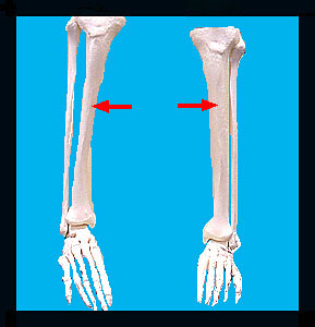
| The lower limb is composed of the proximal femur and distally the tibia, fibula, tarsals, metatarsals, and phalanges. |
 |
 |
| Know the following bones and their specialized features. |
Frontal Bone
Temporal Bone
|